


In the evolving landscape of regenerative medicine, platelet rich fibrin (PRF) is quickly gaining recognition as a powerful biological tool for enhancing healing and restoring damaged tissue. Unlike synthetic materials or pharmaceutical interventions, PRF relies entirely on the patient’s own biology—making it a safe, cost-effective, and increasingly preferred solution across surgica and orthopedic disciplines. Its impact on bone regeneration in particular is turning heads in clinical communities worldwide.
This guest post will explore the PRF treatment process, its role in promoting bone healing, and practical guidance on integrating PRF into regenerative workflows.
What is Platelet Rich Fibrin (PRF)?
Platelet Rich Fibrin is a second-generation autologous blood concentrate obtained without the use of anticoagulants or additives. Upon centrifugation, the blood sample separates into a fibrin-rich matrix that contains a high concentration of platelets, leukocytes, and growth factors.
Unlike platelet rich plasma (PRP), PRF forms a solid, slowly-dissolving scaffold that continuously releases bioactive compounds—offering longer-lasting stimulation of tissue repair and regeneration. It’s a natural biomaterial with strong applications in soft tissue healing, angiogenesis, and crucially, bone regeneration.
Biological Properties That Make PRF Unique
The clinical effectiveness of PRF lies in its structure and content:
- Fibrin Matrix: Forms a three-dimensional scaffold that facilitates cell migration, tissue in-growth, and matrix remodeling.
- Sustained Growth Factor Release: PRF gradually releases TGF-β1, PDGF, VEGF, and other cytokines for up to 10–14 days post-application.
- Leukocyte Inclusion: The presence of white blood cells supports antimicrobial defense and immune-modulated healing.
- Completely Autologous: No additives, making it biocompatible and safe for most patients—even those sensitive to foreign substances.
These properties give PRF a regenerative edge—especially when bone integrity and healing timelines are critical.
PRF Treatment in Bone Regeneration: Clinical Relevance
PRF is used extensively in bone regeneration protocols where promoting osteogenesis, angiogenesis, and tissue integration are key goals. Common clinical applications include:
- Orthopedics: In treating small bone defects, delayed unions, and minimally invasive fracture repair, PRF enhances the healing cascade without introducing foreign graft materials.
- Maxillofacial Reconstruction: Surgeons use PRF membranes to guide tissue regeneration and support bone grafts in facial trauma or tumor resection cases.
PRF works not only as a bioactive additive but often as a primary regenerative agent when combined with particulate grafts or scaffold materials.
PRF vs. Synthetic Grafts: A Biological Advantage
While synthetic bone grafts, allografts, and xenografts have their place in regenerative medicine, PRF presents several advantages:
| Feature | Synthetic Grafts | Platelet Rich Fibrin |
| Biocompatibility | Varies, possible immune reactions | Fully autologous, no rejection risk |
| Osteoinductive | Often limited | High due to sustained growth factor release |
| Handling Properties | Rigid or brittle | Moldable, adaptable to defect site |
| Cost & Accessibility | Can be expensive | In-house preparation from patient’s own blood |
| Infection Risk | Present, especially in xenografts | Minimal due to leukocyte presence |
PRF doesn’t replace all grafting materials but offers a natural alternative or enhancement that can significantly improve patient outcomes, especially in early-stage defects or compromised healing environments.
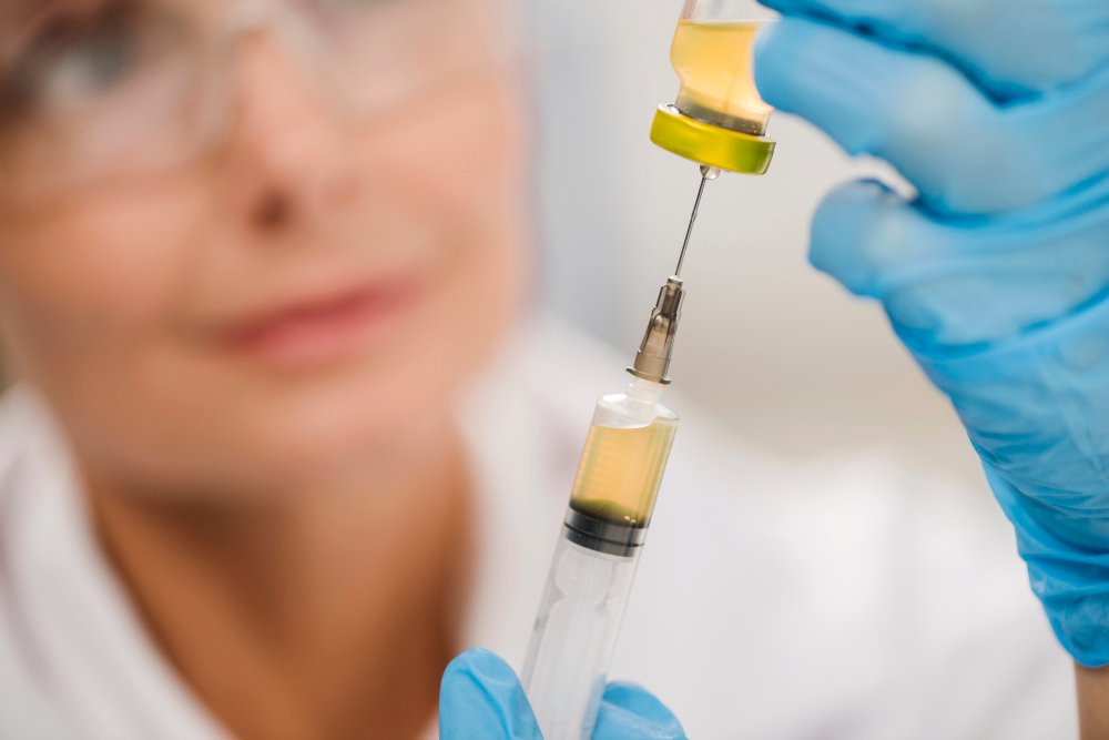
Preparation Protocol: Getting PRF Right
While the PRF preparation process is relatively simple, precision matters. Here’s a step-by-step look at how clinicians typically prepare PRF:
- Blood Collection: Draw 10–20 ml of the patient’s venous blood using sterile PRF tubes.
- Centrifugation: Immediately spin the tubes at optimized speeds (e.g., 2700 RPM for 12 minutes). The absence of anticoagulant ensures natural clotting begins during the spin.
- Fibrin Separation: Post-spin, extract the fibrin clot located between the red blood cell base and acellular plasma top layer.
- Membrane Formation: Compress the fibrin clot in a PRF box to form a membrane suitable for surgical placement or grafting.
Timing is critical. The entire procedure should be performed swiftly (within minutes) to preserve platelet viability and fibrin polymerization quality.
Tips for Effective PRF Use in Bone Regeneration
To ensure the best regenerative outcomes with PRF, consider the following clinical tips:
- Standardize Centrifuge Settings: Use a validated protocol with a medical-grade centrifuge to ensure consistent product quality.
- Combine with Grafts When Needed: PRF can be mixed with bone graft particles to enhance scaffold integration and osteogenic potential.
- Use as Barrier Membrane: In ridge preservation or sinus lifts, PRF membranes function as natural barriers promoting healing while preventing soft tissue invasion.
- Educate the Patient: Patients should understand that PRF is autologous, safe, and enhances healing—improving consent and compliance.
These practices help build a reproducible workflow for incorporating PRF into surgical or orthopedic procedures.
Advancing Bone Healing with Platelet Rich Fibrin: Clinical Perspective
Platelet Rich Fibrin (PRF) has become a cornerstone in bone regeneration protocols due to its natural ability to support both hard and soft tissue healing. Unlike synthetic graft enhancers or pharmaceutical additives, PRF delivers a sustained release of growth factors like PDGF, TGF-β1, and VEGF, which are critical for initiating cellular repair and osteoblast activation.
In clinical practice, PRF has shown to:
- Accelerate bone graft integration during dental implant placement and ridge augmentation.
- Enhance healing in extraction sockets, reducing the risk of dry socket and promoting early bone fill.
- Improve outcomes in maxillofacial surgeries, where PRF membranes serve as biologic scaffolds aiding in both space maintenance and vascularization.
- Reduce healing time and inflammation due to its leukocyte-rich matrix and fibrin-based clot structure.
What sets PRF apart is its completely autologous profile—no additives, no risk of immune reaction, and a protocol that can be performed chairside. Its versatility across specialties continues to expand, offering clinicians a regenerative tool rooted in simplicity and biologic intelligence.
Conclusion: PRF is Shaping the Future of Bone Healing
Platelet rich fibrin is more than a surgical adjunct—it’s a biologically active, easy-to-prepare solution that gives regenerative practitioners a reliable edge in challenging cases. By enhancing natural bone regeneration processes, reducing reliance on synthetic grafts, and supporting faster patient recovery, PRF has earned its place in modern clinical protocols.
Whether you’re an oral surgeon placing implants or an orthopedic specialist treating bone defects, integrating PRF treatment into your workflow could redefine your outcomes—and your patients’ experience.


